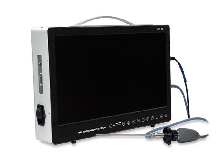
Application of veterinary laparoscope in the collection of bovine oocytes in vitro fertilization.
The recovery rate of oocytes collected by laparoscopic technique is simpler than that of collecting eggs directly from the ovary. In addition, veterinary laparoscopy can be used to check follicular development.
Laparoscope insertion: Insert the blunt trocar into the cannula, insert the cannula into the vagina, position the front end of the cannula at the predetermined position of the vaginal vault through the rectal grasp, then withdraw the blunt trocar, insert the pyramid trocar, and adjust the angle. Make the direction of the trocar and the long axis of the cow slightly to the right at an angle of 10°, and forcefully penetrate the vaginal vault upward. If the resistance decreases suddenly, it indicates that the vaginal vault has been pierced. At this time, take out the trocar. When you hear the sound of air entering and the hand holding the rectum feels the expansion of the abdominal cavity at the same time, it indicates that the piercing position is correct. The stainless steel sheath with the endoscope barrel is inserted into the sleeve. According to the feeling of rectum, if the cavity is small, it needs to be filled with some CO2. The speed of the air intake should generally be controlled at 7~L/min. The amount of gas filled depends on the size of the cow. "Generally, it is Around 9L.
Collection of oocytes: The rectal grasp operator should constantly adjust the position of the laparoscope and the ovary, and display the shape of follicles and corpus luteum at different positions on the ovary, in order to determine the follicles that need to be punctured. An assistant grasps the operator's password and the follicle displayed on the screen according to the rectum, operates the puncture needle, collects the follicular fluid, and puts the follicular fluid into the collection tube that has been added with a small amount of culture fluid. Another assistant is responsible for operating the foot vacuum pump. In order to prevent the loss of oocytes, the vacuum pump should be started (the negative pressure should be 100mm Hg) before the puncture. After collection, the follicular fluid is placed in the test tube rack for 5-10 minutes, and the follicular fluid at the bottom of the collection tube is sucked into the plate with a straw, and then observed under an optical microscope. And the detected oocytes are sucked into the thin tube. The two ends of the thin tube are sealed, and the oocytes can be transported to the laboratory for mature culture. After the operation is completed, the endoscope barrel is taken out, and the two assistants press on both sides of the abdomen wall of the cow to expel the gas in the abdominal cavity. When the gas is no longer discharged, the cannula is taken out and the vagina is checked every other day to prevent infection.
Precautions:
1. Anesthesia: not too shallow or too deep. Effective anesthesia is a prerequisite for the completion of follicular puncture to obtain oocytes. If the anesthesia is too shallow, the cow will feel pain, and it will affect the accuracy of the operation, and the anesthesia should be supplemented. If the anesthesia is too deep, the cow will not stand stably, or even lie down, which will also affect the operation. If the cow is lying down, the operation should be stopped immediately, and the cannula and laparoscopic tube should be withdrawn quickly. At the same time, attention should be paid to release the Baoding in time to avoid injury to the cow.
2. Puncture: The rectal grasper should correctly grasp the puncture angle when puncturing the vaginal vault to prevent puncturing the rectum and large intestine. When puncturing the follicle, the needle should not be pierced too deeply, otherwise it will damage the ovarian substance and cause hemorrhage. This will not only contaminate the follicular fluid, affect the detection of oocytes, block the needles and ducts, but also affect the development of the follicles on the ovary. recover. In case of bleeding, the follicle puncture should be stopped immediately, the needle and catheter and the collection tube should be replaced, and the needle and catheter should be flushed with 0.01mg heparin/ML saline before the next follicle puncture.
3. Follicular fluid collection: The collected follicular fluid should be kept at a constant temperature as much as possible. It is best to detect oocytes immediately after collection, and put them in mature culture medium containing FSH, package them in thin tubes and transport them to the laboratory, generally from The time from harvest to incubator for maturation culture should not exceed 4h.
4. Skills: The successful use of this method requires not only good equipment, but also requires the operator to have a high level of knowledge and proficiency in practical operation, as well as close cooperation with two assistants. Therefore, the necessary training must be carried out before the experimental operation.
In addition, this method can also be used to obtain oocytes from young cows in advance, thereby increasing the utilization period and reproductive potential of the cows, and accelerating genetic progress. This method will be a good way to obtain egg sources after superovulation and the use of isolated ovaries.
