Application of Laparoscopy in Diagnosis and Treatment of Animal Reproductive Diseases
1. In the process of animal embryo transfer, the ovaries and uterus of the donor and recipient should be examined by laparoscopy before the operation. This will not only reduce unnecessary operations, reduce the workload, but also increase the success rate of the operation. Laparoscopy is performed on the recipient before surgery. If there is a normal corpus luteum on the ovary, the embryo will be directly transferred to the uterine horn during the operation. There is no need to check the ovary. This will not only shorten the operation process and reduce the chance of surgical wound infection, but also It is important to avoid mechanical damage to the ovaries and corpus corpus luteum that may be caused by traction and inspection of the ovaries to increase the success rate. The time of laparoscopy should be 1 day before the operation for the donor sheep. Early degeneration of the corpus luteum occurs more frequently in superovulated sheep. If the inspection time is too long from the operation time, the ovarian status may still change, which will affect the accuracy of the diagnosis and reduce the success rate of the operation.
2. Diseases of the ovary
①Early degeneration of the corpus luteum:
Due to the influence of climatic factors or the disorder of hormone secretion, the corpus luteum can degenerate prematurely. 3 to 5 days after the estrus, if the corpus luteum becomes smaller and changes from fleshy red to light yellow, it is the degeneration of the corpus luteum. Embryos cannot be transferred if the recipient's corpus luteum is degraded at an early stage, and embryos may also undergo degenerative changes if the donor's corpus luteum is degenerated at an early stage. The recovery rate of embryos is greatly reduced, or even embryos cannot be recovered at all. Therefore, laparoscopy can be used to observe the changes in the corpus luteum in the ovary, and hormones can be used to prevent the corpus luteum from degenerating prematurely to ensure the recovery rate of embryos.
②Ovarian cyst:
Ovarian cysts in goats treated with natural estrus or superovulation are mostly follicular cysts. The follicle wall is thick, poor in transparency, and grayish white. The number and size of the cysts vary, with small numbers being larger, and multivesicular cysts being relatively small. In goats treated with superovulation, luteal cysts are sometimes seen due to LH or hCG injections during breeding. The corpus luteum cyst slightly protrudes on the surface of the ovary and is dark red with no depression in the center. When ovarian cysts occur, they can be treated by laparoscopic needle puncture.
③ Diseases of the uterus:
When a disease occurs in the uterus, a laparoscope can be used to observe the situation in the uterus, and then some conventional methods can be used for treatment. If uterine effusion occurs, puncture into the uterus with a laparoscopic puncture needle, use a straw to suck out the uterine effusion, and then use drugs to treat it.
When using laparoscopy for disease observation, you should pay attention to the following issues: you cannot check under the condition of being full, otherwise it is not easy to observe, and it is easy to damage the stomach or intestine when inserting a trocar; when inserting a trocar, you must master Good direction and depth, too shallow can not enter the abdominal cavity, too deep or wrong direction will puncture the stomach and intestines; the trocar insertion site should not be close to the spine, otherwise it may damage the kidneys and even cause death; after the trocar is inserted, the lens must be sent in first , Observe whether the feeding position is correct, such as inserting into the mesenteric fat, a large amount of white flashing adipose tissue can be seen. At this time, it is necessary to withdraw and adjust to the front end of the lens in the abdominal cavity and then breathe in. If it penetrates the mesentery, it will stick to the reproductive organs, which will affect the observation, and it is difficult to readjust it. It is necessary to operate in a dust-free room as much as possible to avoid sending dust into the abdominal cavity with the air. After the operation is completed, the air in the abdominal cavity should be released slowly, not too fast, to prevent shock due to the sudden decrease of abdominal pressure and the large amount of blood flowing to the abdominal cavity causing cerebral anemia.
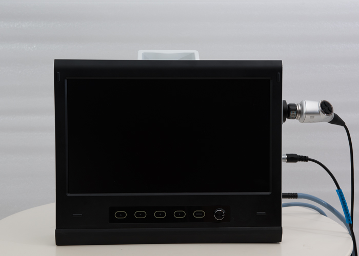
Laparoscopy technology is widely used in the veterinary market, and many large livestock farms have been equipped with veterinary laparoscopes. Xuzhou AKX Electronic Science And Technology Co., Ltd. focuses on the development and production of veterinary endoscopes. Welcome to consult!
