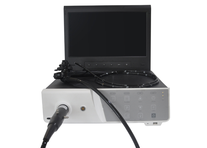
Veterinary electronic gastroscope may be the most commonly used endoscope in the field of small animal endoscopes. It can be used to treat symptoms such as vomiting, salivation, dysphagia, nausea, vomiting, weight loss, anorexia, diarrhea, fecal mucus and tenesmus.

Application of Veterinary Electronic Gastroscope in Dogs and Cats Clinic
1. Indications for veterinary electronic gastroscopy:
Veterinary electronic gastroscope can check many upper gastrointestinal diseases, such as vomiting, nausea, dysphagia, salivation and changes in breath smell. In addition, gastroscopy can be used to check the animal's esophagus and trachea for foreign bodies, or to assess the sensitivity of sick dogs and cats to food. If there is chronic intermittent diarrhea and vomiting, blood in the vomiting, etc., gastroscopy is required.
Veterinary electronic gastroscope can observe irregular mucosa, obtain cytology samples when checking biopsies, and use gastroscope to clarify the deformation and dislocation of different stomach, duodenum and other positions. In fact, many diseases of the stomach and duodenum are on the surface of the mucosa, which can be diagnosed by endoscopy. For upper gastrointestinal diseases, the use of endoscopes for diagnosis can achieve good results, and this method does not cost much.
2. Preparations for veterinary electronic gastroscopy
(1) Preparation for dogs and cats. Before performing veterinary electronic gastroscopy in dogs and cats, fasting and water-free are required to ensure that the stomach and duodenum are empty. If there is food and liquid left in the stomach, it is more difficult to empty the stomach, which will hinder the examination. Most of the gastric mucosa can be seen and can be examined with an electronic gastroscope, but the substance needs to be removed during the examination. If the material is too large or too hard to be removed by aspirator, it is necessary to use biopsy forceps and other equipment to remove it, or gastric lavage to empty the material. If there is barium in the stomach, it usually takes 24 hours to digest, mainly because the barium is easy to stick to the stomach wall, which affects the observation.
(2) Anesthetize dogs and cats. When performing veterinary electronic gastroscopy, in order to ensure the safety of dogs and cats and improve the convenience of operation, they usually need to be anesthetized, and at the same time, electronic gastroscopes and other equipment can be protected. It is necessary to use an opener to avoid sudden awakening of dogs and cats during operation. Avoid using atropine when anesthetizing dogs and cats. Most other anesthetics can be used. Atropine affects the movement rhythm of the pylorus, making it more difficult for the device to pass through the pylorus. Weichangning can be used before anesthesia, which will not affect the movement of the stomach and pylorus. Inhalation anesthetics generally do not affect the operation. Generally speaking, inhalation anesthesia during gastroscopy in dogs and cats is achieved through tracheal intubation, which can avoid the reflux of the contents in the gastrointestinal tract. Moreover, the antiemetic agent metoclopramide will make it more difficult to insert the canine gastroscope catheter, so it needs to be stopped about 15 hours before the gastroscopy.
(3) Baoding for dogs and cats. Generally lying on the left side of Baoding, so that the gastric antrum and pylorus are far away from the table, and the gastroscope can enter the pylorus through angle incision. When using a gastroscope to carry out a gastrostomy catheter operation, you should lie on the right side, so that the gastrostomy tube can smoothly enter the stomach without affecting other abdominal organs.
3. Specific operations and precautions for the application of veterinary electronic gastroscope
If there is abnormality or vomiting in the upper gastrointestinal tract of dogs and cats, the esophagus, stomach and duodenum need to be checked. During the operation, the operator needs to be able to distinguish the tissue and mucosal structure of each segment of the gastrointestinal tract and judge the disorder Condition and get a biopsy from the gastrointestinal tract.
Dogs and cats have been fasted before the gastric examination, there is no food, liquid, or only a small amount of liquid in the stomach, but it is abnormal if there is blood or bile in the stomach. Pay special attention when using a catheter to absorb gastric fluid. Even small fragments of gastric mucosal damage can block the catheter. When inflating the stomach, it is necessary to observe the difficulty of the wrinkles disappearing to determine whether the stomach is swollen. If the stomach swells easily, it indicates that the stomach wall is relatively thick. Then the folds and the appearance of the mucosa are evaluated. Normally, the mucosa is smooth and the color is dark pink or reddish.
(1) Check the connection of the gastroesophagus. Conventional electronic gastroscopy usually evaluates the gastroesophageal junction, which is usually closed for animals. Check the closure of the mucosa at this location, mainly to determine whether there is reflux in the gastroesophagus. Filling with gas can make the wrinkles disappear, which can better observe the mucosa of the esophagus and the gastrointestinal junction, and ensure that the catheter can smoothly enter the animal's stomach. Some animals need to hold the neck esophagus with their hands to avoid dissipating the air that enters it.
(2) Check the gastric body. After the stomach is inserted into the tube, it is necessary to ensure that the tube head of the catheter enters in the center, slightly tilted to the left, and then the catheter is inserted to the right to prevent damage to the esophagus and gastrointestinal junction.
Rotating the catheter can make it easier to observe the gastric body. The right side of the electronic gastroscope is the small gastric curve, the lower left of the fold is the greater curve, and the pyloric antrum and the angle notch are on the upper right. An important criterion for distinguishing the lesser curvature of the stomach and the pyloric antrum is the angle notch. To perform a comprehensive examination of the lesser curvature of the stomach and the pyloric antrum, an examination of the cardia is required. In order to have a comprehensive observation of the lesser curvature of the stomach and the cardia, the gastroscope needs to be bent back so that tumors, foreign bodies, and ulcers will not be missed. This operation is performed in the canine gastric notch and in the midbody of the cat's stomach. These positions allow the inserted catheter to be turned over. In order to see the lesser curvature of the stomach and the cardia, you can turn the internal knob counterclockwise and lift the insertion tube upwards, and inject air into it to ensure that the stomach is expanded, so that the observation field is clear, and the mucosa can be observed. The amount of gas that can be visualized by the Ministry is changing, and careful monitoring is required. The stomach should not be too inflated to avoid affecting the cardiopulmonary function. If the dog or cat is affected by factors such as breathing and heart rate during gastroscopy, it is necessary to use an inhalation device to deflate until the diseased dog and cat is stable. Generally speaking, it is enough to inflate the stomach to separate the folds. If you want to observe the surface of the stomach in a comprehensive way, you need to over-inflate to make the folds disappear and the blood vessels on the mucosal surface can be observed.
(3) Check the pylorus. Through animal electronic gastroscopy, the pyloric antrum and other parts of the stomach are distinguished, and the pylorus and the pyloric antrum are tender enough to show all the peristalsis in the stomach. If the pyloric sinus moves violently, the pylorus will close. Before entering the pyloric sinus, control the insertion tube at the angle notch, and feed the insertion tube into the pyloric sinus up and right. Stomach tumors and gastric ulcers in dogs and cats often appear in the pyloric antrum and lesser curvature of the stomach.
Observation of the pylorus is relatively easy. The pylorus is located at the tail end of the pyloric sinus, but sometimes covered by rain fluid or tissue, the pylorus will be blurred and the catheter will be difficult to pass. To pass the pylorus smoothly, it is necessary to ensure that the pylorus is in the middle of the line of sight. However, it is more difficult for large dogs. Because the catheter is in the dog's mouth, the insertion tube is not very operable. At this time, it is necessary to reduce the amount of gastric inflation and reduce the degree of pylorus and pyloric antrum contraction. The gastroscope cannot be forcibly entered, and the insertion process needs to be patient and slow. The cat’s pylorus is narrower, and it is more difficult to insert the tube through the pylorus. The expansion of the pylorus can cause obvious vagal bradycardia. If this happens, you need to withdraw the insertion tube, deflate appropriately, and wait for the cat to stabilize before operating.
In short, the application of electronic gastroscope in the clinic of dogs and cats can observe irregular mucosa, examine biopsies, obtain samples, and display the anatomical deformation or dislocation of these areas. Although the safety of veterinary electronic gastroscopy is relatively high, it is inevitable that complications will occur. In serious cases, there may even be perforation of the stomach and duodenum, or tears in blood vessels and organs. Therefore, it is necessary to be careful and rigorous in the operation of gastroscopy, to ensure that the inspection of dogs and cats is more smooth and efficient, can timely find problems in the upper digestive tract of dogs and cats, and take effective measures to improve the results of the inspection, and treat dogs and cats more specifically.