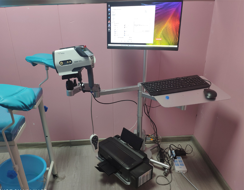
colposcopy has been widely used in the diagnosis of diseases of the lower reproductive system, especially for the pre-cancerous lesions of the lower reproductive tract, early cancer and early diagnosis of sexual diseases.
What is a colposcopy and how is it done?
A colposcopy is used to find cancerous cells or abnormal cells that can become cancerous in the cervix, vagina, or vulva. These abnormal cells are sometimes called “precancerous tissue.” A colposcopy also looks for other health conditions, such as genital warts or noncancerous growths called polyps. A special instrument called a colposcope gives your doctor a lighted, highly magnified view of the tissues that make up your cervix, vagina, and vulva. The colposcope is placed close to the body, but it does not enter the body.
Check steps:
1. The patient takes the bladder lithotomy position, wets the vaginal speculum with normal saline or does not use lubricant, exposes the cervical fornix, and gently wipes off the cervical secretions with a cotton ball.
2. Adjust the height of the colposcopy and examination table for proper inspection, place the lens 10cm away from the vulva (the lens is 15-20cm away from the cervix), aim the lens at the cervix, turn on the light source (using electronic colposcopy, connection number monitor), and adjust The focal length can make the light softer and green filter lens can be added. For more precise blood vessel examination, red filter lens can be added.
3. In order to distinguish between normal and abnormal, squamous epithelium and columnar epithelium, the following solutions can be used:
(1) 3% acetic acid solution (97ml of distilled water + 3ml of pure glacial acetic acid): the columnar epithelium quickly swells, whitish, and changes like grapes. After a few seconds, the junction of phosphorus-columnar epithelium is very clear.
(2) Iodine solution (100ml of distilled water + 1g of iodine + 2g of potassium iodide): color the normal squamous epithelium rich in glycogen to a brown; atypical hyperplasia, cancerous epithelium with less glycogen and no coloration. The columnar epithelium and the epithelium are not stained because of the low estrogen level. The non-stained area is called a positive iodine test.
(3) 40% trichloroacetic acid (60ml of distilled water + 40ml of pure trichloroacetic acid): make the condyloma acuminata appear as needle-like protrusions, with a clear boundary with the normal mucosa.
4. Observe the contents of the cervix size, erosion-like tissue range, whether the cervical mucosa has ectropion; whether the epithelium is abnormal, the range of lesions, the shape of blood vessels, the distance between capillaries, etc.
Attention should be paid to colposcopy:
1. The schedule of colposcopy should generally be arranged 3-5 days after menstruation. There is no time limit for suspected cervical cancer or precancerous lesions. If there are lesions in the cervical canal, it is appropriate to check when it is close to ovulation.
2, 3 days before the examination, stop vaginal douche and medication, and prohibit sex.
3. For those with vaginal inflammation, it is best to check after the inflammation subsides.
