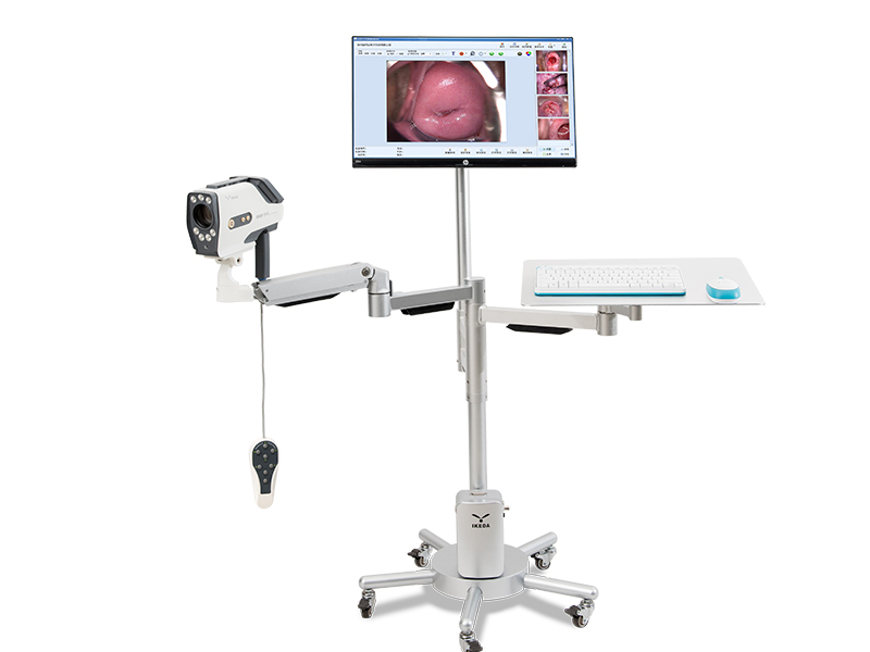
How does the electronic colposcopy work?
1. Clean the product first: the probe should be wiped and disinfected with medical alcohol before and after use.
2. Place the speculum
Do not use any lubricants, such as liquid paraffin, soapy water, etc., when placing the speculum. If the vagina is dry in the elderly, you can apply some saline or a little non-irritating lubricant on the outside of the speculum. Choose different types of speculum according to the patient's age, fat or thinness. When placing the speculum, keep opening the front and back lobes while entering, gradually exposing the cervix. Don't force the speculum directly into the cervix. This can easily damage the cervix and cause bleeding, which affects the observation. Especially for patients with cervical cancer, the speculum should be lighter.
3. Create a new case, and then the system enters the image acquisition window interface. At this time, the image acquisition window will display the dynamic images observed by the colposcopy.
4. Calibrate the image color
Align the colposcope vertically at a distance of about 20cm from the white calibration plate. After the focus is clear, wait 2 seconds for the image to be validated.
5. Zoom in and focus
Adjust the colposcope to a suitable distance from the speculum (10-35CM) until the image on the monitor is clear. When observing local lesions, if local magnification is required, the image is partially magnified. With the gradual magnification of the image magnification, the observer can see a clear image of the cervical cavity or vaginal fornix.
6. Direct observation
By adjusting the focal length, push the lens forward and backward until a clearer image can be observed on the monitor, and then use fine-tuning to make the image very clear, and then you can start to observe. If observing the lesions of the cervix, first use a cotton ball to gently wipe off the secretions on the surface of the cervix and vagina, and then observe. The observation content includes the size of the cervix, whether there is ectropion of the cervical mucosa, the size of the erosion surface, and whether the blood vessels and epithelium are abnormal.
7. Detailed observation
In order to further distinguish the squamous epithelium or columnar epithelium on the surface of the cervix, understand the contractile response of blood vessels, and determine the nature of the lesions on the surface of the cervix, it is sometimes necessary to apply some drugs on the surface of the cervix to make the image clearer and more conducive to the diagnosis. The commonly used methods are as follows:
1) 3% acetic acid solution test: 3% acetic acid solution is the most commonly used solution during colposcopy. After acetic acid is applied to the surface of the cervix, its colposcopy image changes rapidly.
2) Iodine solution test
After exposing the cervix with a speculum, first use a sterile cotton ball to gently wipe off the surface mucus, and then use a small cotton ball dipped in iodine solution to evenly smear the lesion and the surrounding mucosa to observe the local coloration.
3) Trichloroacetic acid solution test
The generally used concentration is 40%-50%, which has a strong corrosion and fixation effect on the organization. Normal cervix or vaginal mucous membranes become white and thickened immediately after applying trichloroacetic acid, but the surface is smooth. Pseudocondyloma (villiform labia minora), the mucous membrane becomes white after applying trichloroacetic acid, and the surface is obviously uneven and rough. Condyloma acuminata immediately showed thorn-like or rod-like protrusions after applying trichloroacetic acid, which is clearly distinguished from the normal mucosa. Trichloroacetic acid has a good therapeutic effect on the early condyloma acuminatum distributed on the mucosal surface. The epithelium of the application site will fall off 2 to 3 days after application, and it can be reused after 1 week.
8. Collect images
After the operator selects the best image observation effect, the current image should be collected in time. By observing the images collected on the monitor, the doctor can explain the lesion to the patient in comparison with the image to achieve a good effect of intuitive communication. In addition, color printout devices can be used to provide color image reports to patients or to facilitate future applications.
9. Generate report
10. Exit the system and shut down
