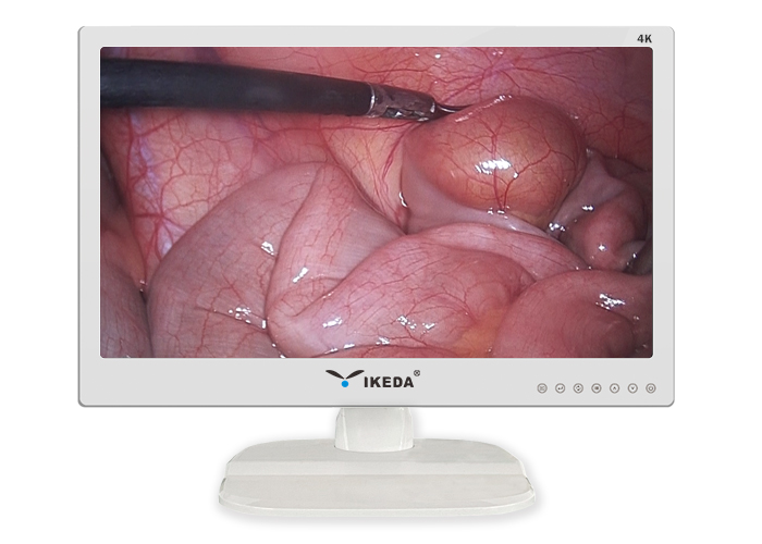
In the digital system, the medical display is the ultimate presenter of medical images. It carries the replacement film, guarantees the image quality, and finally realizes the doctor's "soft reading film" to observe and diagnose the patient, adjust the display correctly, and ensure the working status of the display Ideally, it can fully display the image details. Let's take a look at the debugging method of the medical display.
1. Debug distortion
If the image picture is distorted during the diagnosis, the doctor will be prone to misjudgment, so it is very important to debug the distortion of the professional medical display. You can use professional monitor testing software, or use the OSD menu to debug. First observe whether the four corners of the display and the circular state appearing in the middle are a perfect circle. At the same time, observe whether the square on the screen is square. If not, be patient and debug until the graphics are accurate.
2. Debug brightness and contrast
Because the display's high brightness and contrast can increase the dynamic range of image colors, it helps doctors better judge the light and dark areas of the image. In the process of debugging a professional medical display, the brightness and contrast of the display can be adjusted by setting the light output of the screen to make it look more comfortable, and the dynamic range of the display is larger and clearer.
3. Debug moiré
Because the moiré is an optical interference phenomenon, it will affect the visual effect and cause the diagnosis to be affected to a certain extent, so it is necessary to debug the moiré of professional medical displays. You can mostly select the function of removing moiré in the OSD menu and call up the corresponding menu, and adjust the moiré in both the horizontal and vertical directions until the moiré appears on the display is greatly reduced or even completely eliminated.
4. Debugging focus
The focusing ability reflects the concentration of the electron beam emitted by the picture tube. The image is composed of electron beams that scan across the screen. The focused display can accurately project the electron beams onto the phosphor layer of the display. The bright spots of the display are thin and concentrated, and the images appearing on the screen will not be blurred. . The poor ones are edge divergence. The resulting characters and images are also blurry. You can try to adjust the focus in the OSD menu first. You will often see a noticeable improvement in display quality.
