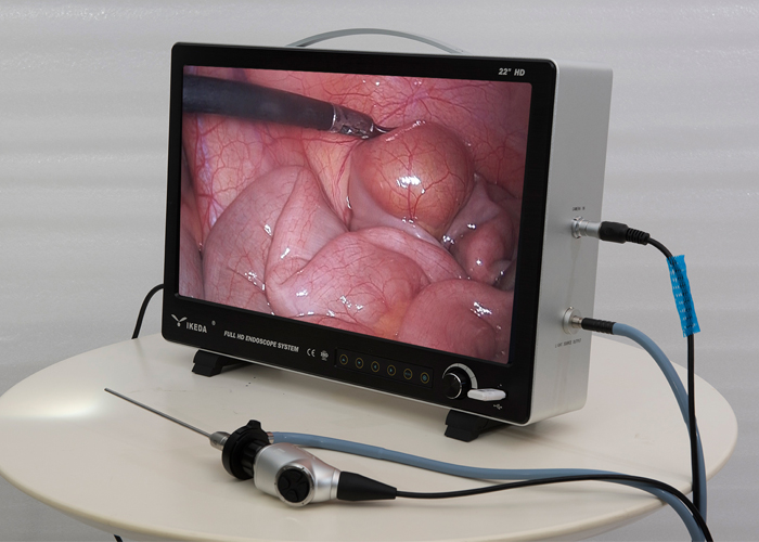
Application of veterinary laparoscope in goat bladder operation
Equipment: general medical CO₂ compressed gas cylinder, surgical laparoscopic system (endoscopic imaging system, monitor, pneumoperitoneum, high-frequency current machine), a set of conventional laparoscopic surgical instruments: cannula: puncture needles, instruments, etc. that enter the abdominal cavity Access; puncture needle: puncture the abdominal wall for other instruments to penetrate; fallopian tube grasping forceps: grasping tissues and organs such as the fallopian tube during the operation; monopolar coagulation forceps: grasping tissue and coagulation to stop bleeding; monopolar coagulation hook: coagulation Hemostasis; Knot pusher: Push the knot into the abdominal cavity through the cannula; Insufflation needle: Connect to the insufflator before the operation to inflate the abdomen.
Animal anesthesia, Baoding, surgical department and pneumoperitoneum preparation: Animals are lying on their backs, and a wide range of hairs are shaved and routinely disinfected from the front edge of the pubic bone to the back of the abdominal bottom wall of the sword cartilage. The surgical department is covered with a sterile isolation towel, ready for surgery.
Use the "three-hole method" to install the cannula and insert the instrument into the abdominal cavity to observe the anatomical structure of the goat's bladder and its surrounding tissues. After the laparoscopic lens enters the abdominal cavity, the outline of the bladder and the distribution of blood vessels on the surface of the bladder can be clearly seen, so that a place with few blood vessels can be selected as the site of bladder incision.

Surgical method of laparoscopic cystectomy and suture: "three-hole method" puncture cannula
The surgeon and assistant use a pair of wound pliers to clamp the skin on both sides of the umbilicus, and lift them upwards. The surgeon holds a pneumoperitoneum to puncture the abdominal cavity vertically from the umbilicus. After checking to confirm that the insufflator needle enters the abdominal cavity, connect to the insufflator through the insufflator needle. Set the pneumoperitoneum pressure value of 1.06kPa, and inflate the abdomen at an airflow rate of 1L/min. When the set pneumoperitoneum pressure is reached, turn off the pneumoperitoneum machine and withdraw the pneumoperitoneum needle.
The surgeon and assistant use the wound pliers to clamp the skin on both sides of the umbilical hole and lift it up. The surgeon uses a 10mm diameter cannula with a puncture needle to vertically puncture into the abdominal cavity, pull out the puncture needle, and connect the pneumoperitoneum through the cannula. Turn on the cold light source and imaging system, insert the laparoscopic lens from the sleeve, and observe the goat’s bladder through the monitor. The other two puncture cannulas with a diameter of 5 mm were punctured from the anterior edge of the ilium into the abdominal cavity.
Make a traction thread on both sides of the planned incision of the bladder: a triangular needle threaded through the silk thread is sent into the abdominal cavity through the entire layer of the abdominal wall. With the aid of grasping forceps, use needle-holding forceps to hold the triangular needle, and pass the serosa muscle layer on the side of the planned incision of the bladder to make a traction line; The thread tail is knotted outside the body and fixed outside the abdominal wall.
Incision and suture: Use surgical scissors to cut the bladder wall at the predetermined incision and enlarge the incision to be about 5cm long, and thoroughly flush the bladder through the bladder catheter. Use a suction device to suck out the flushing fluid in the pelvic cavity. After the incision is cleaned up, with the aid of grasping forceps, use the needle-holding forceps to hold the medical needle-absorbable suture to make a layer of nodule to suture the serosa-muscular layer of the bladder wall. After the suture is completed, enter the irrigation tube of the suction device from the sleeve to flush the suture of the bladder incision.
Treatment of the puncture hole of the abdominal wall cannula: After the operation is completed, close the pneumoperitoneum, withdraw the lens, and pull out the cannula. The skin puncture hole with the diameter of 10mm cannula is sutured with a needle, and the skin puncture hole with the diameter of 5mm cannula is not sutured. Apply tincture of iodine to the puncture hole. Continuous intramuscular injection of antibacterial drugs 3 days after surgery. The nodular suture at the puncture hole of the body surface skin was removed 10 days after the operation.
When performing laparoscopic surgery, with the help of the endoscope imaging system, the surgeon can directly view the local anatomy of the abdominal organs, and with the magnification of the laparoscopic, the surgeon can clearly determine the operation site and perform the operation. The operation caused little tissue damage, the incision and sutures healed well, and there was no adhesion between the operation site and other organs. The animals recovered quickly after the operation.