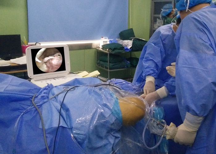
Application Of Arthroscopy In The Lysis Of Knee Joint Adhesions
Surgical methods:
Epidural or spinal anesthesia, tourniquet on the base of the thigh. Water is punctured above the knee to continuously expand the joint cavity. Incisions of approximately 0.6 cm in length are made on the inside and outside of the knee as the entrance for arthroscopy and instrument operation. If the adhesion in the joint cavity is severe and it is difficult to enter the arthroscopy, first use the self-made loosening hook to enter the joint cavity on the outside of the knee. When the knee joint is straightened between the patellofemoral joint and in the suprapatellar capsule, it is repeatedly fan-shaped and blunt. Sexual separation. At this time, a large amount of adhesion tissue may be brought out by the hook and create a space, and then placed in an arthroscopy for observation. Use a nucleus pulposus clamp or planer to remove the adhesion tissue. Check the intercondylar fossa and both sides of the chamber, the anterior cruciate ligament and the medial and lateral meniscus in turn, remove and clean the adhesions and hyperplastic synovium. If there are meniscus injuries, osteophytes, loose bodies, etc., they can be treated under arthroscopy. If the adhesion between the chambers on both sides is serious, it can be used to loosen the probe hook, blunt or sharp peeling with a hook knife. Move the knee joint. If the increase in flexion is not satisfactory, the lateral patellar retinaculum can be loosened under arthroscopy; an incision of about 1.5 cm is made at the inner and outer edges of the suprapatellar rectus femoris, and a stripper is placed in the midfemoris muscle and femur. Blunt peeling between the bones, use a knife to cut or plan a knife to remove the medial femoral muscle and surrounding adhesion tissue. Use continuous force to flex the knee joint. At this time, you can hear the sound of the adhesion band breaking in the joint. If the knee joint mobility is still unsatisfactory, use a stripper to separate the expansion of the quadriceps muscle and the internal and external femoral condyle adhesions to loosen, and continue to separate the intermediate femoris until the knee joint flexes more than 120°. Passiveness is required. There is no resistance and no rebound force during flexion. After the operation, a negative pressure suction was placed in the joint, the knee was bent at 90°, and a large cotton pad was compression bandaged. The quadriceps muscle was contracted 24 hours after the operation, the dressing was changed 2 to 3 days, the negative pressure drainage blood volume was less than 50ml/24h and removed, and the knees were bent autonomously and CPM was exercised.

Knee joint adhesion is mainly caused by adhesion, contracture, and fibrosis of the knee joint sliding device caused by lower limb bone and joint trauma, surgery, long-term knee joint immobilization, intra-articular injury or inflammation. After femoral shaft fractures, it is easy to cause adhesions of the quadriceps muscles, especially the scar contracture and fibrosis of the intermediate femoris muscle, which seriously affect the flexion and extension of the knee joint. After long-term immobilization, the patellofemoral joint adhesion, the suprapatellar capsule disappeared, the lateral supporting ligaments of the patella and the joint capsule adhered, and the patella could not move. At the same time, the articular cartilage also undergoes degenerative changes. Arthroscopic technology to loosen and remove intra-articular adhesions and scars; for contracture adhesions around joints, use small incisions, blunt and sharp peeling methods to extend contracture tissues, and release adhesions with less trauma; arthroscopy Loosen the medial and lateral patellar support bands, which is easy to operate and less damage; for those with longer stiffness, peeling off the adhesions between the internal and external condyles of the femur can effectively solve the contracture; small incisions on both sides of the suprapatellar rectus femoris tendon to avoid damage to the thigh The quadriceps combined tendon and its muscle tissue structure provide conditions for early active exercise of muscle tissue and joint accessory structure; peeling between the intermediary femoris muscle and the femoral shaft periosteum can be used to transect or partially remove the proliferative periosteum and contracture fibrosis The intermediary femoris muscle can remove the adhesion tissue and can also improve the elasticity of the rectus femoris and medial and lateral femoris muscles; this operation is a minimally invasive operation, which can completely loosen the adhesions while protecting the normal tissue structure as much as possible, which is conducive to flexion and extension of the knee joint as soon as possible. Get down to exercise.
Principles of arthroscopy technique application:
①From the inside to the outside: remove the hyperplastic adhesion tissue and synovium filling the patellofemoral joint space and the suprapatellar capsule, remove the contracture joint capsule and adhesion zone; plan to remove the hyperplasia synovium, fat pad and adhesion tissue in the intercondylar fossa and both sides of the chamber, Restore the joint space. At this time, the joint range of motion can be improved. At the same time, check the damage of the anterior cruciate ligament, the medial and lateral meniscus, and the articular cartilage. Sharp dissection, resection of the midfemoris muscle, etc. When there is an internal fixation, the inside of the joint should be taken out first, and the outside of the joint should not be taken out.
②Blunt-sharp combination: Try to peel off bluntly in each step, and then loosen sharply under the microscope to remove the scar adhesion tissue.
③ Manipulative cooperation: The operation of loosening and manual manipulation alternately can avoid unnecessary surgical damage and complications such as avulsion fracture or patellar ligament rupture.
The application of arthroscopy to treat knee joint adhesions has the advantages of less trauma, thorough release, early functional exercise, fewer complications, and faster functional recovery. It can also check and treat other intra-articular diseases.