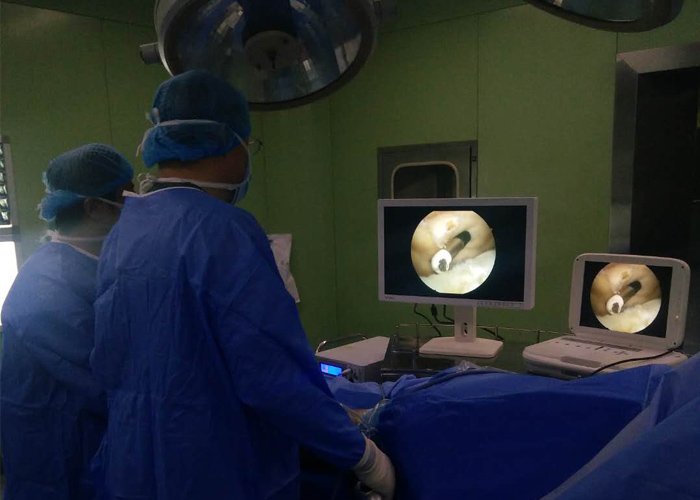
Surgical method: continuous epidural anesthesia or spinal anesthesia is used for the operation. Conventional airbag tourniquet on the base of the thigh. Insert the large water injection tube into the suprapatellar capsule, and enter the joint through the anterolateral, anteromedial, or central entrance of the patellar ligament through the arthroscopy. First, flush each compartment with normal saline until it becomes clear, then remove blood clots and other impurities such as bone cartilage fragments in the joints, and then perform arthroscopy. During the operation, first check the meniscus and ligament damage, check the compression degree of the fracture surface, the size of the fracture, and the fracture displacement. The joint damage is often more serious than that seen on the X-ray film. I-II degree collateral ligament and cruciate ligament injuries are treated non-surgically, III-degree collateral ligament injuries are repaired after platform fracture surgery, and III-degree cruciate ligament injuries are reconstructed after the second-stage fracture has healed. Meniscus injuries are generally peripheral tears, barrel-handle cracks or valvular fissures. According to the injury, partial resection, subtotal resection, and repair and suture are performed. Clear the blood clots between the fractures and insert the soft tissues. The fracture reduction can be under the monitoring of arthroscopy, through the joint, through the fracture line or opening the window to pry or push the top of the collapsed fracture. After the articular surface is reduced and leveled, Kirschner wires are used to temporarily fix the fracture according to the condition, and the bone defect cavity is filled with cancellous bone for bone grafting. After the reduction was satisfied under X-ray fluoroscopy, it was fixed with cancellous bone screws or combined with portal nails. However, if the fracture is still unstable after internal fixation and axial load is desired, an external fixator should be used to support and fix it according to the injury. Also, if the split fracture is larger, a small incision outside the joint capsule should be made and a supporting plate should be used for internal fixation.
Surgery points:
① Thorough debridement is required during the operation. Use spatula, planer, nucleus forceps and other instruments to debride the synovial membrane, blood clot, and osteochondral mass at the fracture end, so as to ensure the accurate reduction of the fracture.
②The fracture line should be explored and the center of the fracture collapse should be found accurately. Sometimes the meniscus should be hooked up to see the entire fracture line or the center of the collapse. For example, for Hohl type II fractures, find the center and lowest point of the collapse under the microscope, locate it and drill Kirschner wires with the aid of X-ray fluoroscopy, and then drill the Kirschner wires with a trephine, so as to accurately open the fenestration, reduction and implantation. bone.
③For the reduction of split-displaced fractures, you can use longitudinal traction, lateral traction, and middle squeeze to reset while prying and pulling under the microscope. During prying and pulling reduction, a hollow nail guide pin can be inserted into a larger bone block to pry out. After the articular surface is flat, the guide pin is further driven and the hollow nail is screwed into the guide pin for compression fixation.
④Bone grafting is emphasized in collapsed fractures and the amount must be sufficient, so as to ensure the articular surface is flat and prevent later collapse.

The advantages of arthroscopic technology in tibial plateau fractures:
① Arthroscopy is the most effective method to determine the actual size of the articular surface dislocation, which can ensure a good reduction of tibial plateau fractures. Use arthroscopy to directly observe the fracture to ensure that the articular surface is flat, and instruments such as probes can be used to assist in the reduction of the bone mass, and to remove the embedded soft tissue, bone fragments and broken cartilage.
②The application of arthroscopy can check the soft tissues for combined injuries and deal with them as appropriate to improve the integrity of the surgical plan. Arthroscopy directly prompts a good intra-articular field of vision, and can directly observe the intra-articular structure, avoiding the limitation of the naked eye.
③Under the arthroscopy, you can directly observe whether the screw enters the joint cavity, and guide the direction of the screw and the degree of tightness after screwing.
④ Arthroscopic surgery has been carried out in saline irrigation to remove blood clots and bone cartilage fragments thoroughly, and the sterile environment is good, which avoids the exposure of articular cartilage and other tissues to the air and reduces the chance of infection.
⑤ Arthroscopy is a minimally invasive technique that can appropriately expand the indications for surgery. For example, the tibial plateau hohl type I fracture with obvious joint swelling, the function of arthroscopy is to clear the blood in the joint and check and treat the ligament or meniscus injury, so as to reduce the traumatic reaction and reduce the intra-articular adhesion.
⑥ Arthroscopic surgery is less dependent on skin and other soft tissue conditions, with small surgical incisions, less interference with blood supply, and strong tissue healing ability, which can avoid exposure of steel plates and bones.